The medical sector, especially pathology, is one of our key areas of interest. Hyperspectral imaging enables the development of innovative techniques, such as new methods for diagnosing cancer. This allows for faster and more cost-effective diagnoses, speeding up surgeries and reducing the time patients need to remain under anesthesia while awaiting the pathologist’s results.
Since our cameras cannot be calibrated before the optics are mounted, adapting them to a microscope was possible but rather complicated. The camera needed to be mounted to the microscope, and the final calibration had to be conducted on-site, which often meant that the calibration conditions were not ideal.
Development of a Relay Optics
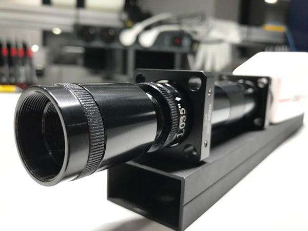
By developing a relay lens for the hyperspectral FireflEYE, we have overcome this disadvantage. The camera and the relay lens are assembled at our facilities, where they are directly calibrated according to our high-quality standards.
With the relay lens extension, we provide full flexibility to our customers, allowing them to work with the camera (plug-and-play). Whether it is mounted on a microscope, used with one of our common lenses with different focal lengths, or even attached to any specialized optics with a C-mount.
Adding close-up lenses
After further design studies, we found that using a conventional lens in combination with close-up lenses, also from Schneider-Kreuznach, was effective. We used the lens with the smallest available field of view, namely the 50 mm lens with a 6.5° field of view. With a mounting adapter on the 50 mm lens, a close-up lens can be added, which directly leads to a spot size in the millimeter to centimeter range.
The advantage of this solution for the customer is significant. It is now possible to individually add various close-up lenses to meet specific requirements for spot size on a macroscopic scale. The accuracy of the camera is not affected, as it is calibrated in our lab with the 50 mm lens, and the transmission of the close-up lenses is nearly 100%.
To demonstrate how easy it is to work with the camera, we attached the relay lens to the C-mount adapter of a Zeiss Axiophot, which was provided by the ILM (Institute of Laser Technologies in Medicine and Measurement Technique), a research institute in Ulm. From the start – mounting the camera to the microscope – to the first measurement, it took only a few minutes, and we obtained a sharp image with a richness of detail.
Obtaining reflectance values is even easier than in other measurement environments. By simply using the microscope in light transmission mode, rather than having the light source illuminate the sample from above, the camera can be white-calibrated by taking an image without a sample placed on it. As the light passes through the sample, the transmitted signal carries the relevant information due to particular absorption features within the sample. With this setup, we achieve excellent results with an integration time of no more than 0.1ms.
Histological Analysis
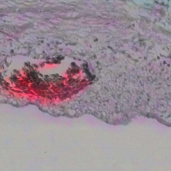
We analyzed a histological slice of pig skin cultured on a chicken egg. Under the microscope, the sample exhibits a complex structure with various colors. The figure below shows a colored infrared (CIR) image of the pigskin histology on the left-hand side, while on the right-hand side, the spectral reflectance of all pixels inside the corresponding rectangles with the same color in the image can be seen. Since the Axiophot has an NIR filter for wavelengths greater than 750nm, the spectral range is limited to this value.
Visualizing hidden Details
The different colors result from the dyeing methods applied to help visualize various features of the histological sample: coagulated areas due to the laser cut, healthy skin, hair follicles, blood vessels, and erythrocytes.
Analyzing the hyperspectral image of the samples allows the user to extract hidden features, visualize them using different band settings in an RGB output, and even quantify the results automatically, for example, with the help of machine learning. Below are additional examples of histological measurements taken with the hyperspectral FireflEYE 185.


About the Author
Dr. Matthias Locherer has been the Sales Director at Cubert GmbH since 2017. With a PhD in Earth Observation from Ludwig Maximilian University of Munich, he brings extensive expertise in remote sensing, spectral imaging, and data analysis. Matthias has contributed to numerous research projects and publications, particularly in the hyperspectral monitoring of biophysical and biochemical parameters using hyperspectral satellite missions. His deep knowledge of optical measurement techniques and physical modeling makes him a key driver in advancing innovative hyperspectral technologies at Cubert.


